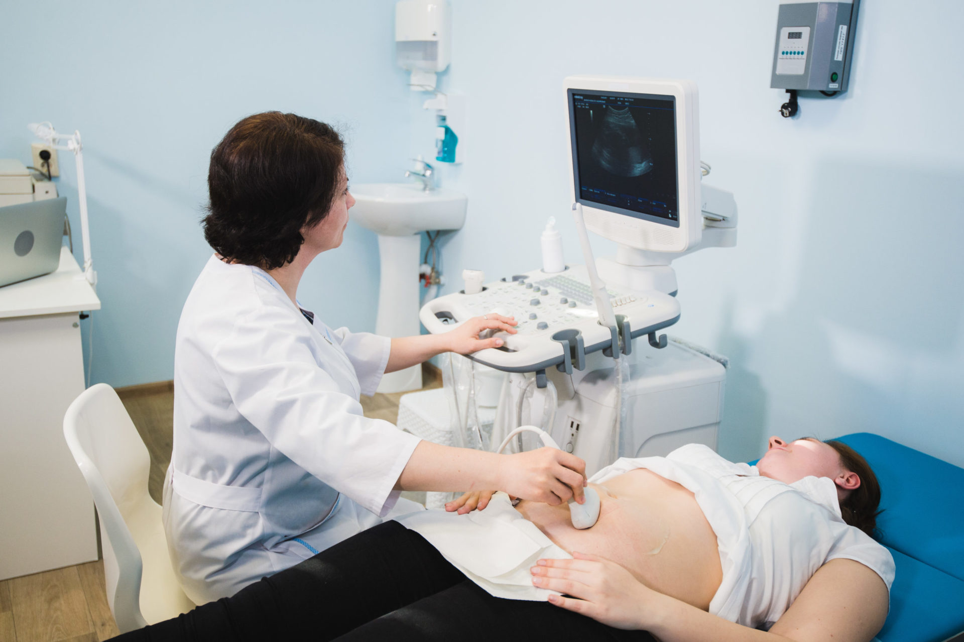
One of the more common fetal anomalies I encounter is enlargement of the baby’s kidneys. This is called fetal hydronephrosis. (Hydro—water and “nephro” meaning kidney). It is most commonly detected at the routine 20 week anatomy ultrasound scan we perform. This may occur in as many as 4% of pregnancies. This renal enlargement typically resolves and is of little concern after delivery, especially if the dilation is mild. Sometimes the enlargement is even more transient and may resolve as the pregnancy progresses.
For some perspective, at the time of the 20 week anatomy scan, your baby’s kidneys are typically less than 4mm. If the renal enlargement is 4 to 10 mm in size, it is considered to be “mild” dilation. If greater than 10mm in dilation, there is increased concern for persistent renal abnormalities that may require intervention shortly after delivery.
For my patients with mild hydronephrosis, (greater than 4 mm), at 20 weeks, I generally repeat the US at 28 weeks, in the third trimester. If the renal measurements are still enlarged, (greater than 7), I refer my patients to our pediatric urologist for consultation. THIS IS NOT FOR INTERVENTION IN THE PREGNANCY, but simply to establish care for follow up after delivery, if needed at that time. After delivery pediatricians typically repeat an ultrasound 1 week later and have the baby follow up with the pediatric urologist if necessary.
WHAT COULD BE WRONG?
It is very important to note that a large proportion of babies with measurements between 7 and 10 are ultimately normal and not associated with other abnormalities. The dilation resolves, if not by delivery, then shortly thereafter.
For those without resolution, there may be an anatomical abnormality involving the baby’s renal system. There may be an obstruction; this may occur between the kidney and the bladder or from the bladder through the urethra, which is the tube from the bladder providing elimination. There may also be insufficiency or inappropriate action of the valve between the bladder and the ureter, (the tube which carries urine from the kidney to the bladder), causing reflux. These possibilities will be thoroughly evaluated, if needed, by the pediatric urologist after delivery.
Fetal hydronephrosis has a very loose association with Down’s syndrome, in that as many of 18% of Down’s babies may have this abnormality. Fortunately, patients may have already had some form of genetic screening, prior to the 20 week anatomy scan, that may relieve anxiety. Genetic screening may be considered for patients who have not had any evaluation.


