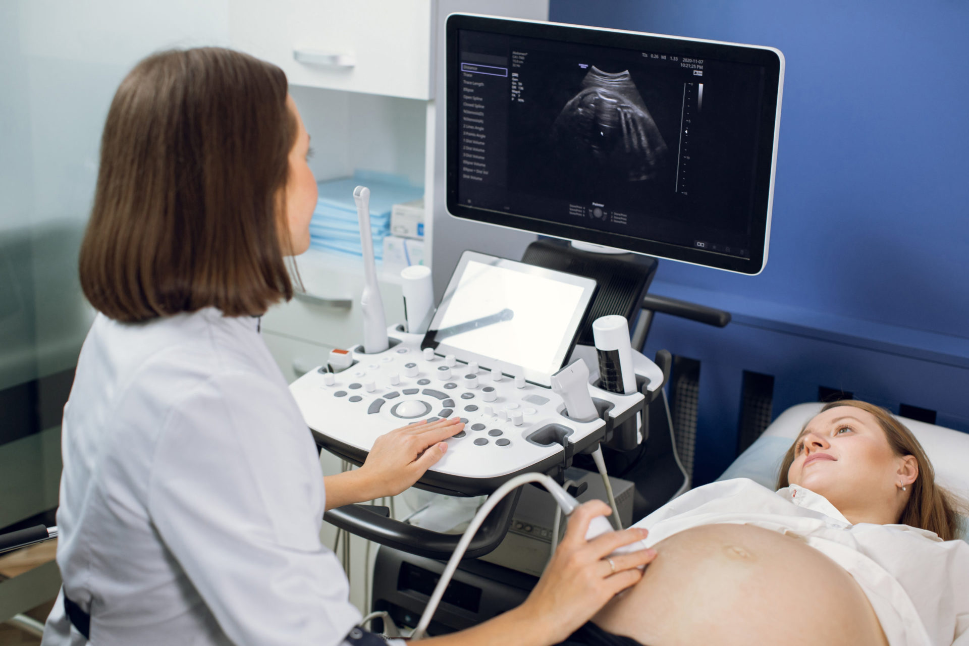
Measurement of fetal nuchal translucency is an optional ultrasound you may have been offered. The nuchal translucency is a measurement of the back of the baby’s neck, and if the baby has chromosomal abnormalities or particular congenital defects, this measurement may be thickened. Typically performed between 12-14 weeks of gestation, this is a screening test for potential chromosomal anomalies, in particular Down syndrome. However, there are other concerns that may be associated with an abnormal nuchal translucency. This thickening may also be seen in pregnancies associated with congenital heart defects.
Nuchal translucency measurement is a key component of the First-Trimester screening test. It is important to understand that for patients who intend to perform cell-free fetal DNA testing, nuchal translucency may not be offered or discussed. For patients pursuing cell-free DNA testing, this ultrasound is relevant only in a very narrow application—that of potentially identifying congenital defects.
Because the nuchal translucency measurement is traditionally associated with the first-trimester screening test, (and detection of chromosomal abnormalities), confusion arises when offered with cell-free fetal DNA testing. I find that nuchal translucency is beneficial for screening congenital defects, in particular, cardiac anomalies. Strictly speaking, this is a very narrow application of the nuchal translucency test and it will by no means rule out all cardiac defects. However, I have experienced patients with significant cardiac anomalies revealed, indirectly, by abnormal nuchal translucency, despite normal chromosomal testing with cell-free fetal DNA. In these rare cases, patients were able to make significant clinical decisions far earlier than would otherwise be possible, as cardiac anomalies are typically discovered much later, at the anatomy scan offered at 20 weeks.